A Novel Sox-based Continuous and Homogeneous Assay for the Discovery of Inhibitors of Inactive and Active AKT
Susan Cornell-Kennon1, Matthew Hakar1, Hayley McMahon1, Daniel Urul1, Erik Schaefer1, Earl May1
1. AssayQuant Technologies, Inc., 260 Cedar Hill Street Marlborough MA 01752 United States of America

Abstract
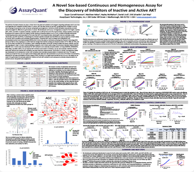
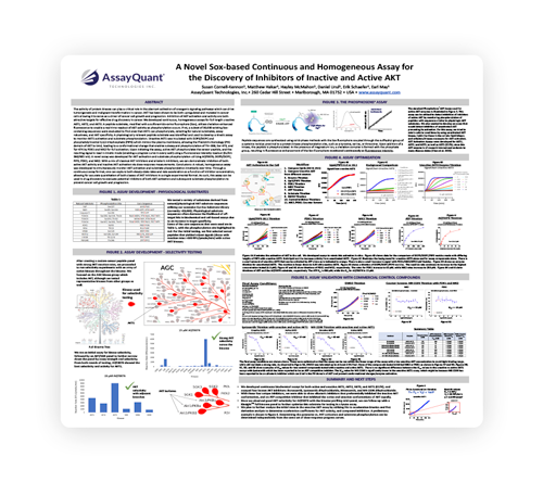
The activity of protein kinases can play a critical role in the aberrant activation of oncogenic signaling pathways which can drive tumorigenesis and malignant transformation in cancer. AKT has been shown to be both upregulated and mutated in cancer cells allowing it to serve as a driver of cancer cell growth and progression. Inhibition of AKT activation and activity are both attractive targets for effective drug discovery in cancer. We developed continuous, homogeneous assays for full length unactive AKT1, AKT2, and AKT3. A peptide substrate, modified with a sulfonamido-oxine fluorophore (Sox), utilizes chelation-enhanced fluorescence to enable a real-time readout of AKT activity as phosphorylation occurs.
First, a subset of 30,000 existing Soxcontaining sequences were evaluated to find ones that AKT1 can phosphorylate, selecting for natural substrates, assay robustness, and AKT specificity. A physiologically relevant peptide substrate was identified and used to develop a kinetic assay to monitor AKT1 activation and substrate phosphorylation. Unactive AKT1 was incubated with DOPS/DOPC and phosphatidylinositol 3,4,5-trisphosphate (PIP3) which mimics the plasma membrane, allowing the Pleckstrin Homology (PH) domain of AKT to bind, leading to a conformational change that enables subsequent phosphorylation of Thr-308, Ser-473, and Tyr-474 by PDK1 and MK2 for full activation. Upon initiating the assay, active AKT phosphorylates the sensor peptide, and the resulting signal is read in kinetic mode (enabling a progress curve in every well) using a fluorescence intensity readout (Ex/Em 360/485 nm). A novel assay was developed for AKT activation and substrate phosphorylation utilizing AQT0076, DOPS/DOPC, PIP3, PDK1, and MK2. With a mix of classical AKT inhibitors and allosteric inhibitors, we can demonstrate inhibition of both active AKT activity and inactive AKT activation via dose-response measurements.
Conclusions: A robust, homogeneous assay was developed to simultaneously monitor AKT activation and substrate phosphorylation kinetically over time. Through a continuous assay format, one can capture both steady-state rates and rate acceleration as a function of inhibitor concentration, allowing for accurate quantitation of both classes of AKT inhibitors in a single experimental format. As such, this assay can be used in drug discovery to evaluate potential inhibitors of both AKT activation and subsequent substrate phosphorylation to prevent cancer cell growth and progression.
Key Takeaways
- We developed continuous biochemical assays for both active and unactive AKT1, AKT2, AKT3, and AKT1 (E17K), and assayed four known AKT inhibitors: Borussertib, Ipatasertib dihydrochloride, Miransertib, and MK-2206 dihydrochloride.
- With these four unique inhibitors, we were able to show allosteric inhibitors that preferentially inhibited the inactive AKT conformation, and an ATP competitive inhibitor that inhibited the active and unactive conformations of AKT equally.
- Since we observed good AKT selectivity for AQT0076 with the kinome profiling mini-panel, we can follow-up with a KinsightTM full kinome panel to further optimize this substrate for testing in a lysate assay.
- We plan to further analyze the initial rates in the unactive AKT assay by utilizing fits to acceleration kinetics and first derivative analysis to determine acceleration coefficients for AKT activity, and compound inhibition. A preliminary example is shown in Figure 6. Determining the potencies vs. AKT activation and substrate phosphorylation can be determined independently from the same set of dose-response progress curves.
